
How OCT Imaging Helps Monitor Glaucoma Progression
Glaucoma is a progressive eye condition that can cause permanent vision loss if not carefully monitored. Because it often develops without noticeable symptoms, many patients are unaware of changes happening within their eyes. Early detection and precise tracking are essential to protecting long-term vision. At DeNovo Eye, advanced technology plays a key role in diagnosing glaucoma early and monitoring its progression with precision and confidence.
Understanding Glaucoma and Why Monitoring Matters
Glaucoma is a group of eye conditions that damage the optic nerve, typically due to increased intraocular pressure (IOP). The optic nerve is responsible for transmitting visual information from the eye to the brain. Once nerve fibers are damaged, they cannot regenerate.
The goal of glaucoma care is not to reverse damage, but to slow or stop further progression. That requires:
• Early detection
• Accurate baseline measurements
• Ongoing monitoring
• Timely treatment adjustments
This is where OCT imaging makes a significant difference.
What Is OCT Imaging?
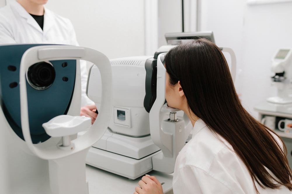
Optical Coherence Tomography (OCT) is a non-invasive imaging technology that uses light waves to create detailed cross-sectional images of the retina and optic nerve. It allows your eye doctor to see the microscopic layers of the retina and measure the thickness of nerve fiber tissue with incredible accuracy.
How OCT Detects Glaucoma Damage
Glaucoma primarily affects the retinal nerve fiber layer (RNFL) and the ganglion cell layer - both critical components of your visual system.
OCT imaging helps by:
• Measuring Nerve Fiber Thickness: One of the earliest signs of glaucoma is thinning of the retinal nerve fiber layer. OCT can detect this thinning before noticeable vision loss occurs. By comparing your measurements to a normative database and to your own previous scans, your doctor can identify subtle changes over time.
• Evaluating the Optic Nerve Head: OCT provides detailed images of the optic nerve head, including the cup-to-disc ratio. Changes in this structure can indicate progression of glaucoma.
• Establishing a Baseline: When glaucoma is suspected or newly diagnosed, an initial OCT scan establishes a baseline measurement. Future scans are compared to this baseline to detect progression - even if changes are very small.
• Tracking Progression Over Time: OCT software can analyze trends across multiple visits. This helps determine whether nerve fiber loss is stable or worsening, allowing for timely treatment adjustments.
The Importance of Routine Eye Exams
Glaucoma is a chronic condition that requires regular monitoring to protect your vision over time. Even when eye pressure appears stable, subtle structural changes can still occur within the optic nerve. Because these changes often happen without noticeable symptoms, routine eye exams are essential. Staying consistent with regular eye exams is one of the most important steps you can take to preserve your sight and maintain lifelong visual health.
Protect Your Vision with Advanced Glaucoma Monitoring
Glaucoma management is no longer based solely on eye pressure readings and visual field tests. OCT imaging has transformed how we detect and monitor this condition by allowing eye doctors to see microscopic structural changes in the optic nerve before significant vision loss occurs.
If you have glaucoma, elevated eye pressure, or a family history of the condition, don’t wait for symptoms to appear. Schedule your next eye exam at DeNovo Eye and learn how advanced imaging can help safeguard your sight. Visit our office in McKinney, Texas, or call (469) 317-2020 today.


MAP
Powered by: 